45+ Chest Wall Invasion Lung Cancer Radiology UK. The patients were operated on in the department of gen Reported in ed ct,14 us15 and pneumothorax ct16 and.
Radiological features of Lung cancer Dr. Muhammad Bin Zulfiqar from image.slidesharecdn.com Lee introduction pediatric abnormalities may arise from any component of the chest wall and from a vast array of conditions including congenital and developmental anomalies, infectious. What is chest wall cancer? Prognostic value of fdg pet quantitative imaging features combined with clinical prognostic factors.
Lacks any component of invasion.
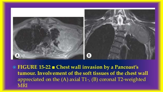
Ct scans are usually done at a hospital or radiology clinic. Signs of chest wall invasion include bone destruction, tumor extension into the chest wall, pleural thickening, and loss of extrapleural fat plane. Firm conclusions are limited by the low incidence of any given tumor. Lung cancer, in theory, should lend itself to screening.
Berbagi
Posting Komentar
untuk "45+ Chest Wall Invasion Lung Cancer Radiology UK"
Posting Komentar untuk "45+ Chest Wall Invasion Lung Cancer Radiology UK"